Home›Blog ›Collimator radiography for X-ray machines
Collimator radiography for X-ray machines
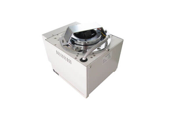
The collimator radiography is used to limit X rays, and Newheek collimator radiography has developed its own medical image enhancement software that improves image quality in all directions. All images are processed in multi-frequency domain, which can realize the separation of different image organizational structures, clear image organizational levels, and perfect combination of well-designed image processing algorithm and high-performance computing library, making it possible to display excellent images in real time. X-ray machine adopts multi-frequency domain noise reduction algorithm to suppress the noise in different frequency domain. The image is delicate, soft and clear. Image correction, image filtering and other rich functions can be realized.The dynamic acquisition of collimator radiography greatly increases the information content of images. Imaging with a large area of 17 x 17 inches can meet all clinical requirements. It has whole body splicing technology. The feature points of the overlapping images are matched and fused seamlessly, making the transition of the Mosaic image smoother and more natural.
LED manual collimator radiography, fully intelligent field setting, light soft and low energy consumption. Extra long life free of maintenance.One key switch machine control box to simplify the process of power on and off, and avoid human factors fault.
Newheek collimator radiography is divided into manual, electric and automatic. The manual collimator radiography is divided into three models: NK102, NK103 and NK202.Stationary and mobile X-ray machines usually use NK102 collimator radiography. The maximum exposure field of collimator radiography is 440mmx440mm (SID=100cm), and the single lighting time of the light field lamp is 30S, meeting different demands.
Author:Glinda
X Ray Collimator
Blog
- Latin American Rural Hospital – Reliable Beam Control for Fluoroscopy
- European Private Clinic – Cost-Effective Upgrade for Mobile Radiography
- Newheek Portable DR System: Integrated Mobile Stand & NK102 Collimator Case Study
- Newheek NK102 Collimator Case Study: Improved Patient Safety & Diagnostic Efficiency at a Regional Hospital
- Veterinary X-Ray Table: How Collimators Elevate Pet Imaging Safety & Accuracy
contact us
TEL:+86 19015366638
E-mail:newheekcn@163.com
Company:Weifang Newheek Electronic Tech Co., Ltd.
ADD:E Building of Future Star Scientific Innovation Industrial Zone of No.957 Wolong East Street, Yulong Community, Xincheng Sub-District Office, Weifang Hi-tech Zone, Shandong Province, China