Home›Blog ›How does the collimation process contribute to dose optimization in medical imaging?
How does the collimation process contribute to dose optimization in medical imaging?
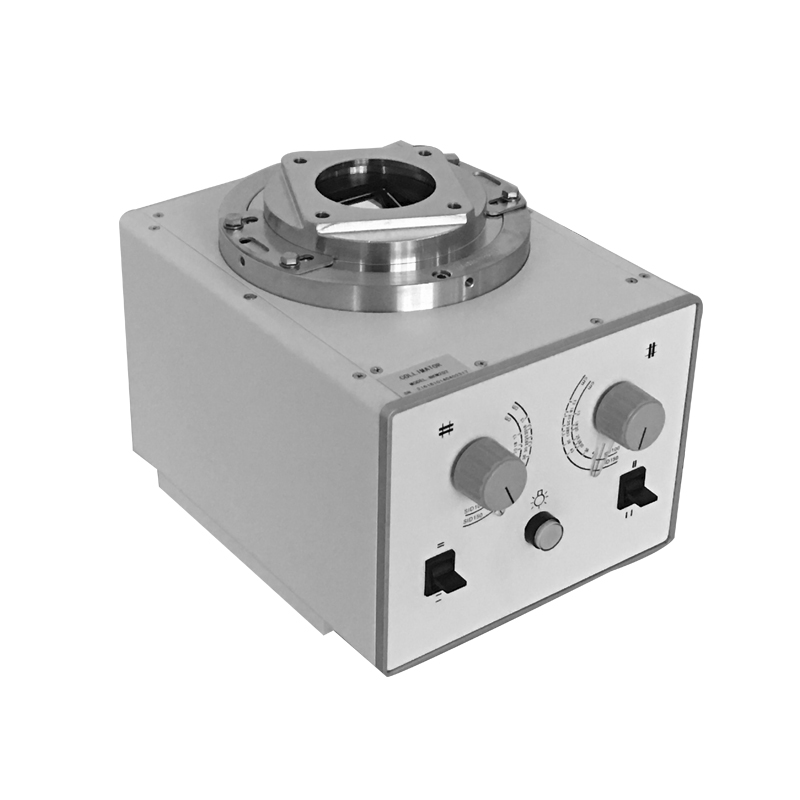
Collimation in medical imaging refers to the process of restricting the size and shape of the X-ray beam to the area of interest, typically the anatomy being examined. This is achieved using collimators, which are devices that can shape and limit the X-ray beam. The collimation process plays a crucial role in dose optimization in medical imaging for several reasons:
1. Reduced Radiation Exposure to Surrounding Tissues:
– By collimating the X-ray beam to only the area of interest, unnecessary exposure to surrounding healthy tissues is minimized. This is particularly important in situations where adjacent structures or organs are not relevant to the diagnostic question. Restricting the X-ray beam helps to focus the radiation dose where it is needed, reducing the overall dose to the patient.
2. Improved Image Quality:
– Collimation helps to improve image quality by reducing scatter radiation. Scatter radiation occurs when X-rays interact with tissues and change direction. If the X-ray beam extends beyond the area of interest, it can contribute to scattered radiation, degrading image quality. Collimation minimizes the spread of the X-ray beam, resulting in clearer and more detailed images.
3. Compliance with ALARA Principles:
– ALARA stands for “As Low As Reasonably Achievable,” and it is a radiation safety principle aimed at minimizing radiation exposure to patients and healthcare providers. Collimation is a practical application of the ALARA principle, as it allows medical professionals to tailor the X-ray beam to the specific needs of the examination, avoiding unnecessary radiation exposure.
4. Reduction of Artifact Formation:
– Collimation helps to reduce artifacts in medical images. Artifacts are unwanted features or distortions in an image that can be caused by various factors, including scatter radiation. By limiting the X-ray beam to the area of interest, collimation minimizes the likelihood of artifacts, leading to more accurate and reliable diagnostic information.
5. Meeting Regulatory Requirements:
– Many regulatory bodies and organizations establish guidelines and standards for radiation safety in medical imaging. Proper collimation is often a requirement to comply with these standards. Compliance helps ensure that healthcare facilities are using best practices to minimize radiation exposure and optimize patient safety.
In summary, the collimation process in medical imaging contributes to dose optimization by focusing the X-ray beam on the specific area of interest, thereby reducing unnecessary radiation exposure to surrounding tissues, improving image quality, and aligning with radiation safety principles and regulatory requirements. This not only enhances patient safety but also supports the goal of obtaining diagnostically valuable images with the lowest possible radiation dose.
Welcome to contact us.
Email :service@newheek.com
Tel:+86 18953679166 ; Whatsapp:+86 18953679166
Author:X Ray Collimator
X Ray Collimator
Blog
- Newheek NK103 C-Arm Collimator Transforms Clinics in Nigeria & Kenya
- Newheek NK103 X-ray Collimator for C-arm in Brazil & Mexico
- NK202X X-Ray Collimator: How It Improves Safety and Clarity in Real-World Use
- NK202X X-Ray Collimator: The Core Component for Safe, Precise X-Ray Imaging
- Latin American Rural Hospital – Reliable Beam Control for Fluoroscopy
contact us
TEL:+86 19015366638
E-mail:newheekcn@163.com
Company:Weifang Newheek Electronic Tech Co., Ltd.
ADD:E Building of Future Star Scientific Innovation Industrial Zone of No.957 Wolong East Street, Yulong Community, Xincheng Sub-District Office, Weifang Hi-tech Zone, Shandong Province, China